
*This page contains men’s anatomy photos.*
We understand that accurate diagnosis is the foundation of successful sports hernia treatment. Our comprehensive physical examination process combines years of specialized experience with meticulous attention to detail. We take the time to thoroughly assess each patient's condition, identifying the subtle signs that often go undetected elsewhere. Whether you're a professional athlete or weekend warrior, our diagnostic approach focuses on pinpointing the exact cause of your groin pain so we can develop the most effective treatment plan for your specific situation.
Our examination methodology integrates clinical expertise with a thorough understanding of athletic biomechanics to distinguish sports hernias from other conditions with similar symptoms. Together, we'll work to uncover the root cause of your discomfort and chart a clear path toward recovery and return to performance.
Recognizing Sports Hernias: Key Clinical Indicators
When three or more of these clinical signs are present, a sports hernia diagnosis can be confidently established.
Pinpoint tenderness at the pubic tubercle where the conjoint tendon attaches
Pain when the deep inguinal ring area is examined through gentle palpation
Discomfort or dilation of the external ring without a detectable hernia mass
Sensitivity or pain at the base of the adductor longus tendon when examined
Generalized aching pain throughout the groin that may extend to the perineum, inner thigh, or across the midline
Advanced imaging techniques such as MRI, CT scan, and ultrasound may be employed to visualize potential tears in the oblique muscles. However, these tests often present interpretation challenges and frequently yield false-negative results. Rather than confirming a sports hernia, imaging is typically more valuable for identifying alternative causes of groin pain, such as hip-related injuries.
A comprehensive physical examination is essential and should thoroughly evaluate all relevant anatomical structures: the abdominal wall, groin region, surrounding nerves, pelvic structures, adductor tendon complex, hip joint, and gender-specific anatomy (the spermatic cord and testicles in male patients, and the round ligament in female patients).
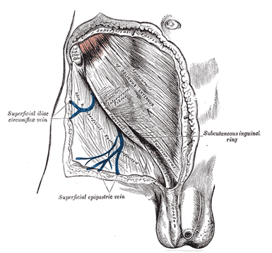
A patient with a Sports Hernia will have an enlarged external inguinal ring secondary to a tear of the external oblique fascia. The external ring can be palpated by inverting the scrotum with the finger. Usually, only the tip of the finger can be inserted into the external inguinal ring. In cases of a Sports Hernia, a finger easily passes through the external inguinal ring. The palpation of the external inguinal ring will often reproduce the patient’s pain. Often the floor of the inguinal canal can be palpated, and the thickness and strength have to be compared to the contralateral side. A thin and weak inguinal floor is also characteristic of a Sports Hernia.

Another essential part of the examination is palpation of the external oblique as it runs parallel to the inguinal ligament. Often a defect can be palpated. A defect in the external oblique fascia is consistent with a Sports Hernia.


.png)
.png)